 by
by Brain MRI Basics: Understanding Common Neurological Findings and Scan Results
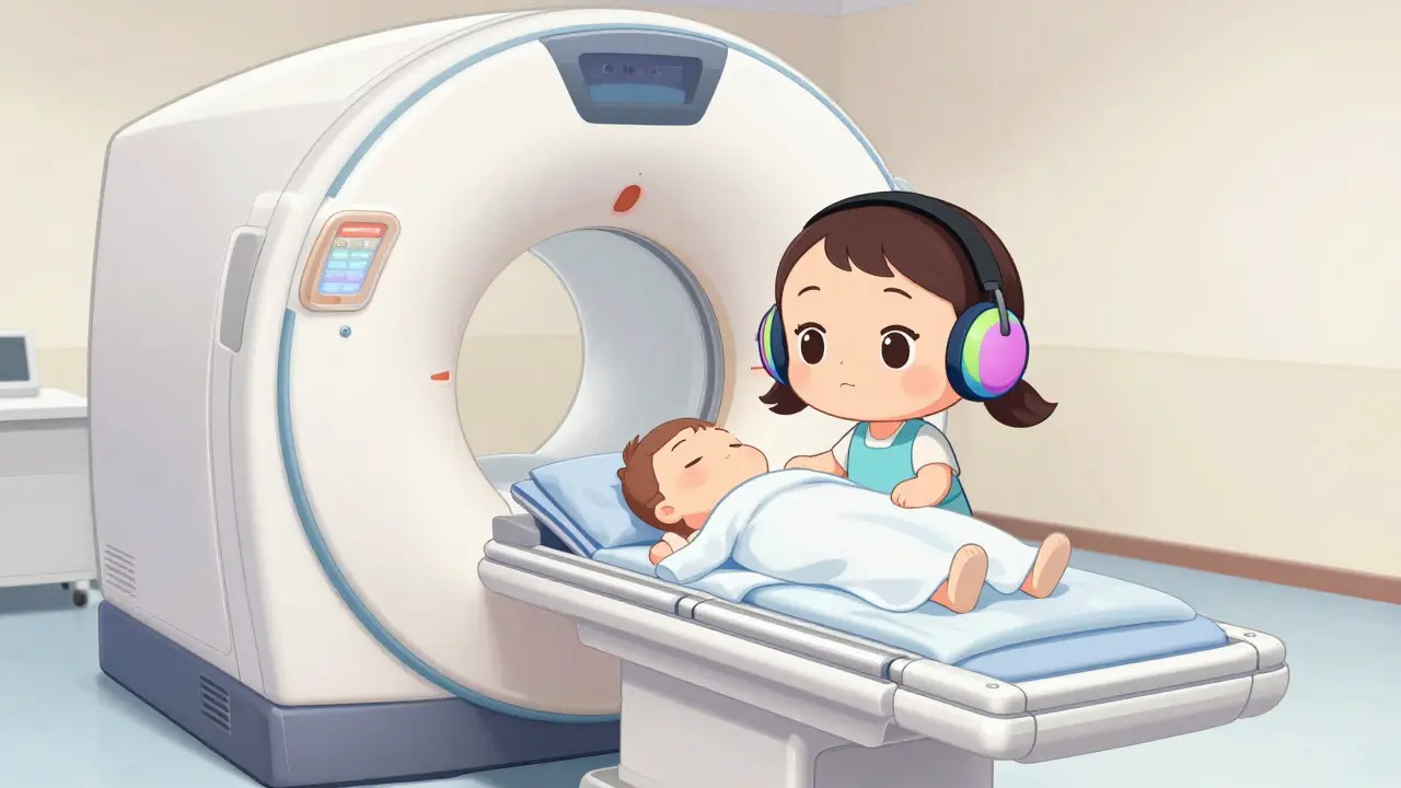
25 Mar, 2026Imagine lying in a narrow tube while a machine makes loud knocking sounds. You stay perfectly still for half an hour. Afterward, you wait days for a report filled with medical terms you don't understand. This is the reality for many people facing a brain scan. You might wonder what the doctor actually sees on those black-and-white images. Understanding the basics of a Brain MRI is a non-invasive diagnostic technique that uses powerful magnetic fields and radio waves to generate detailed cross-sectional images of brain structures without ionizing radiation can help you navigate this process with confidence. This guide breaks down the technology, the sequences used, and the common findings radiologists look for.
What Is a Brain MRI and How Does It Work?
When you hear the term magnetic resonance imaging, it sounds like science fiction. In reality, it is a standard tool in modern medicine. The machine uses a strong magnetic field to align the hydrogen atoms in your body. Radio waves then knock these atoms out of alignment. When the waves stop, the atoms return to their original position, emitting signals that a computer captures to build an image. Unlike a CT scan, this process does not use X-rays or ionizing radiation. This makes it safer for repeated monitoring of chronic conditions.
The technology has evolved significantly since the 1970s. Today, most hospitals use systems with magnetic field strengths of 1.5T or 3.0T. The number refers to Tesla, a unit of magnetic field strength. A 3.0T machine provides about 40% higher signal-to-noise ratio than a 1.5T system. This extra clarity helps doctors visualize small structures like cranial nerves. The scan typically takes between 30 to 45 minutes. You must remain still because any movement can blur the images, similar to taking a photo with a shaky hand.
Understanding the Different MRI Sequences
A single MRI scan isn't just one picture. It is a collection of different images called sequences. Each sequence highlights different tissue properties. Radiologists use these variations to spot specific problems. Here are the most common ones you might see in a report.
- T1-weighted imaging: This sequence shows anatomy clearly. Fat-containing structures appear bright white, while cerebrospinal fluid (CSF) looks dark. It provides the best definition of the brain's physical structure.
- T2-weighted imaging: In this view, water-rich structures appear bright. This makes edema, or swelling, stand out. However, because CSF is also bright, it can sometimes be hard to tell the difference between fluid and pathology without other sequences.
- FLAIR (Fluid-Attenuated Inversion Recovery): This is a special T2 sequence that suppresses the CSF signal. The fluid turns dark, but the pathology stays bright. It is crucial for detecting lesions near the ventricles, which is common in multiple sclerosis.
- DWI (Diffusion-weighted imaging): This sequence detects the movement of water molecules. It is the most important tool for diagnosing acute stroke. It can identify changes within minutes of onset, whereas other methods might take hours.
- SWI (Susceptibility-weighted imaging): This sequence is highly sensitive to blood products. It can reveal microhemorrhages that are too small to see on standard images.
Dr. David Van Der Weide, a Professor of Neurology, notes that T2-weighted and FLAIR sequences are indispensable for detecting most brain lesions. However, he warns they cannot distinguish lesions from CSF without FLAIR suppression. This is why a complete protocol includes multiple types of images.
| Sequence Type | Fluid Appearance | Primary Diagnostic Use |
|---|---|---|
| T1-weighted | Dark | Anatomical definition, fat detection |
| T2-weighted | Bright | Edema, inflammation, general pathology |
| FLAIR | Dark (suppressed) | Multiple sclerosis plaques, periventricular lesions |
| DWI | Bright (restricted) | Acute ischemic stroke detection |

Brain MRI Versus CT Scan: Which Is Better?
Patients often ask why they need an MRI when a CT scan is faster. Both are imaging tools, but they serve different purposes. A CT scan uses X-rays and is much quicker, usually completed within five minutes. This speed is vital in emergency trauma situations where a patient is unstable. However, CT scans expose you to radiation and have lower soft tissue contrast.
Brain MRI offers superior soft tissue contrast resolution, approximately 100 times better than CT for differentiating gray and white matter. This detail allows doctors to see subtle abnormalities that CT might miss. For example, MRI is better at visualizing the posterior fossa, the lower back part of the brain, where CT suffers from artifacts caused by skull bone. According to recent data, MRI detects multiple sclerosis plaques with 97% sensitivity compared to 65% for CT. It also finds small acoustic neuromas as small as 2mm, while CT typically requires tumors to be 5mm or larger.
Cost and accessibility are factors to consider. A brain MRI costs significantly more, ranging from $1,200 to $3,500 per study in the U.S., compared to $500 to $1,500 for a CT scan. Not every hospital has an MRI machine; about 98% of hospitals have CT capability, but only 76% have MRI. Despite the cost, the Royal Australian College of General Practitioners considers MRI the gold standard for investigating most central nervous system diseases.
Common Neurological Findings on an MRI
When a radiologist reviews your scan, they follow a systematic approach. They start with midline structures like the ventricles and move outward to the brain lobes. They are looking for specific patterns that indicate disease. Here are some of the most common findings you might encounter in a report.
Ischemic Stroke: If you have had a stroke, the DWI sequence will show bright areas where water diffusion is restricted. An ADC value below 600 x 10^-6 mm^2/s indicates acute infarction. This finding changes management in 85% of suspected stroke cases, allowing for rapid treatment decisions.
Multiple Sclerosis: This condition affects the protective covering of nerves. On a FLAIR image, radiologists look for bright spots called plaques, often located near the ventricles. Distinguishing these from normal aging changes is critical. Normal periventricular hyperintensities are present in 15% of patients under 50 but 90% of those over 70. Location matters; temporal lobe involvement might suggest herpes encephalitis, while parietal-occipital patterns could indicate posterior reversible encephalopathy syndrome.
Brain Atrophy: As we age, the brain naturally shrinks. Radiologists evaluate cerebral atrophy on FLAIR images rather than T2-weighted ones to avoid overestimation. On T2 images, bright CSF can make the ventricles look larger than they are. Significant atrophy can indicate dementia or Alzheimer's disease, which is expected to affect 12.7 million Americans by 2050.
Tumors and Lesions: MRI can detect acoustic neuromas in the cerebellopontine angle. Dr. Michael Hoch, a radiologist, notes that checking this area in every patient occasionally detects incidental small tumors. These might be vestibular schwannomas that are too small to cause symptoms yet.

Safety, Limitations, and Patient Experience
While MRI is safe for most people, it is not without limitations. The strong magnetic field means you cannot bring metal objects into the room. Patients with certain implants, like pacemakers or cochlear implants, may not be eligible for the scan. You must inform the technologist about any metal in your body, including shrapnel or surgical clips. Safety training is required for all personnel annually under Joint Commission standards.
Another challenge is the noise. The machine makes loud banging sounds as the gradients switch on and off. Technicians provide earplugs or headphones to protect your hearing. Claustrophobia is also a common issue. Some patients find the narrow tunnel of the scanner too confining. Open MRI machines exist but often provide lower image quality. Sedation might be an option if anxiety is severe.
Time is another factor. If you are in a traumatic emergency, MRI is often not the first choice because it takes too long. CT is preferred for acute trauma to rule out skull fractures or immediate bleeding. MRI cannot reliably distinguish the age of a lesion without contrast enhancement, whereas CT shows clear temporal evolution of hemorrhage appearance. Despite these hurdles, MRI remains the dominant tool for neurological imaging through 2035.
Preparing for Your Scan
To get the best results, preparation is key. You will likely be asked to remove all metal jewelry, watches, and hairpins. Some facilities require you to wear a hospital gown. If you have a tattoo with metallic ink, tell the staff, as it can sometimes cause heating or distortion. Eating and drinking are usually permitted unless you are having a scan with contrast dye. Contrast agents improve the visibility of blood vessels and tumors but carry a small risk of allergic reaction.
During the scan, the technologist will talk to you through an intercom. They will tell you when to hold your breath or stay still. If you feel uncomfortable, you can use the call button provided. Remember, the goal is to get clear images so your doctor can make an accurate diagnosis. Understanding the process reduces anxiety and helps you stay cooperative during the 30 to 45 minutes in the machine.
Is a brain MRI painful?
No, the procedure itself is painless. You lie still on a table that slides into the scanner. The only discomfort might come from the noise or the tight space, which is why ear protection is provided.
How long does it take to get MRI results?
Results are usually reviewed by a radiologist within 24 to 48 hours. However, your doctor may take a few days to discuss the findings with you in person.
Can I have an MRI if I have a pacemaker?
Most traditional pacemakers are contraindications for MRI due to the magnetic field. However, newer MRI-conditional devices may be safe. You must consult your cardiologist and the imaging center beforehand.
Why is MRI better than CT for brain scans?
MRI provides much better soft tissue contrast without radiation. It detects subtle abnormalities like multiple sclerosis plaques or small strokes that CT scans often miss.
What do bright spots on a brain MRI mean?
Bright spots can indicate various things, including small strokes, inflammation, or normal aging changes. A radiologist interprets these based on location and sequence type to determine the cause.
Knowledge empowers you when dealing with medical tests. By understanding the basics of how a brain MRI works and what the findings mean, you can have more productive conversations with your healthcare provider. Whether you are monitoring a chronic condition or investigating new symptoms, this imaging technology provides a window into your brain's health that no other tool can match.
Raphael Schwartz
March 27, 2026 AT 07:29mri machines are too expensive for regular folks
Jefferson Moratin
March 27, 2026 AT 10:19The philosophical implications of being able to visualize the mind's physical substrate are profound. When we consider that consciousness emerges from these neural structures we can now image with such precision, we must question what it means to be human. The MRI represents a bridge between the material and the immaterial, between what we can measure and what we experience subjectively. This technology forces us to confront the reality that our thoughts, memories, and emotions have a physical location that can be mapped and analyzed. It is both empowering and unsettling to know that the essence of our being can be captured in grayscale images.
Agbogla Bischof
March 27, 2026 AT 12:46Great explanation of the different sequences! I work in radiology and I can confirm that FLAIR is absolutely essential for MS diagnosis. The DWI sequence is what we use most in stroke cases because it shows changes within minutes. Many patients don't understand why they need multiple sequences but each one provides different information that's critical for accurate diagnosis. The 3.0T machines do provide better resolution which helps with small lesions.
Stephen Alabi
March 28, 2026 AT 20:50This article presents a rather superficial understanding of MRI technology. The claim that MRI is the gold standard is debatable when one considers the cost-benefit ratio for certain conditions. CT scans remain superior for acute trauma situations despite the radiation exposure. The article fails to adequately address the limitations of MRI in emergency settings where time is critical. Furthermore, the assertion that MRI will remain dominant through 2035 is speculative at best given the rapid advancement of other imaging modalities.
peter vencken
March 29, 2026 AT 09:58I had my first brain mri last year and it was way less scary than i expected. The tech guys were super nice and kept talking to me through the intercom. They gave me earplugs for the noise which really helped. The whole thing took about 40 mins but time flew by. Knowing what to expect beforehand made a huge difference for me.
Kevin Y.
March 30, 2026 AT 08:47This is an exceptionally well-researched article that provides valuable information for anyone facing a brain scan. I appreciate the detailed breakdown of the different MRI sequences and their specific applications. The comparison table is particularly helpful for understanding when each type is most useful. The safety information is crucial and should be shared more widely. I would recommend this resource to anyone who needs to undergo neurological imaging.
Aaron Sims
April 1, 2026 AT 02:58And who's funding all these expensive MRI machines? Big Pharma wants you sick and dependent on their treatments. They need you to get scans that find 'abnormalities' that don't really matter. The whole medical-industrial complex is designed to keep you afraid and spending money. Wake up people!
Mihir Patel
April 2, 2026 AT 08:36OMG I had the worst experience with claustrophobia during my mri!!! The machine was so loud and i felt like i was going to die in there. They tried to give me sedation but i was already too scared. My doctor said i needed to come back another time but i dont think i can go through that again. The noise was like a million hammers hitting my ears and the space was so tight. I literally started crying halfway through and they had to stop the scan. Now im so anxious about needing another one for followup. This is the worst medical experience ever!
Blessing Ogboso
April 3, 2026 AT 02:01I want to share that having an MRI can be a deeply transformative experience when you approach it with the right mindset. Understanding that this technology exists to help us better understand our health and wellbeing is important. I've worked with many patients who initially felt afraid but found that education about the process helped them feel more empowered. The key is to remember that you are not alone in this experience and that healthcare providers genuinely want to help you. When we approach medical procedures with curiosity rather than fear, we often find that the experience is less intimidating than we expected. It's also wonderful to see how medical technology continues to advance and improve our ability to diagnose and treat conditions.
Caroline Dennis
April 4, 2026 AT 00:43FLAIR sequence suppression of CSF signal is critical for periventricular lesion detection. DWI ADC values below 600 indicate acute infarction. T1 provides superior anatomical definition.
Marissa Staples
April 5, 2026 AT 06:03I think it's really interesting how much technology has changed medical imaging. My grandmother had a CT scan in the 90s and it was so different from what people get now. I wonder if in another 30 years we'll have even better ways to look at the brain without machines. Maybe something that doesn't require lying in a tube at all. The thought of being able to understand our own brains better is both exciting and a little scary. I guess that's true for a lot of medical advances though.
Kevin Siewe
April 6, 2026 AT 10:29If you're feeling anxious about an upcoming MRI, remember that you can always ask for sedation if needed. The technologists are trained to help patients through the process. Don't hesitate to communicate any concerns you have before the scan starts. Many facilities now have open MRI options for those with claustrophobia. Your comfort and safety are priorities for the medical team.
Chris Crosson
April 7, 2026 AT 13:37I'm curious about the contrast agents mentioned. How common are allergic reactions really? I've heard they can be problematic for some people but the article says it's a small risk. Would love to know more about what the actual numbers are for contrast reactions.
Katie Putbrese
April 8, 2026 AT 02:02People need to stop making excuses for not getting proper medical care. If you can't handle a 45 minute scan, that's a weakness. The article clearly states MRI is the gold standard and yet people here are complaining about noise and claustrophobia. Get over it and prioritize your health. American healthcare is already expensive enough without people wasting resources on unnecessary scans because they're too scared. If you can't handle basic medical procedures, maybe you shouldn't expect the healthcare system to accommodate every fear.