
 by
by Pneumothorax Symptoms and Emergency Care: What You Need to Know
6 Feb, 2026A collapsed lung-medically called pneumothorax-isn't just a scary term. It's a real emergency that can strike suddenly, even in healthy people. If you feel sharp chest pain and can't catch your breath, time is critical. This article explains what to watch for, when to act, and how doctors treat this condition using the latest medical guidelines.
What exactly is pneumothorax?
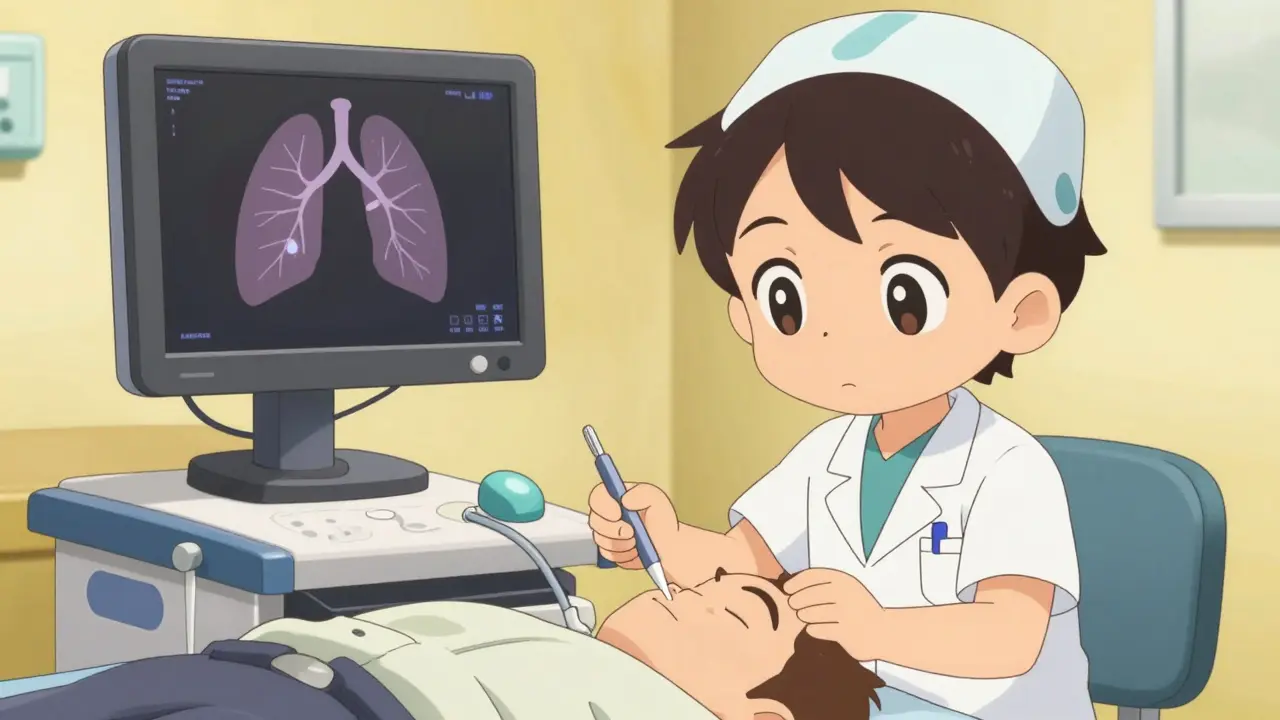
When air leaks into the space between your lung and chest wall-called the pleural space-it creates pressure that stops the lung from expanding properly during breathing. This is a medical emergency. The American Thoracic Society's 2023 guidelines state that pneumothorax affects 7.4 to 18 per 100,000 people annually for primary cases (no lung disease), but rates jump significantly for people with underlying lung issues like COPD or cystic fibrosis. The condition has four main types: primary spontaneous (healthy lungs), secondary spontaneous (with lung disease), traumatic (from injury), and iatrogenic (from medical procedures like chest surgery).
Key symptoms of a collapsed lung
Your body sends clear signals when pneumothorax happens. Here's what to watch for:
- Sharp chest pain that gets worse when you breathe deeply or cough. This pain often travels to your shoulder-a sign noticed in 92% of clinical cases.
- Shortness of breath in 85-92% of cases. If your oxygen saturation drops below 90%, it's an emergency.
- Rapid heart rate over 134 beats per minute. This is a critical warning sign for tension pneumothorax.
- Blue lips or skin (cyanosis) due to low oxygen levels.
- Decreased breath sounds on one side of your chest when a doctor listens with a stethoscope.
These symptoms aren't always obvious. For small collapses (less than 15% lung involvement), you might only feel breathless during exercise. But if you have more than 30% collapse, you'll likely struggle to breathe even at rest. The American College of Chest Physicians found that symptoms worsen rapidly-each minute counts when dealing with this condition.
When to call 911 immediately
Not all pneumothorax cases are equal. A tension pneumothoraxa life-threatening complication where air builds up under pressure, shifting organs and blocking blood flow can develop within minutes. This requires immediate emergency action. Call 911 if you experience:
- Severe difficulty breathing or inability to speak full sentences
- Low blood pressure (systolic below 90 mmHg)
- Tracheal deviation (windpipe pushed to one side)
- Heart rate over 134 beats per minute with cold, clammy skin
The Eastern Association for the Surgery of Trauma's 2022 position paper emphasizes that tension pneumothorax is a clinical diagnosis. Don't wait for X-rays if you have these symptoms-every second counts. In the U.S., emergency departments following best practices achieve diagnosis-to-treatment times of just 8 minutes for tension cases.
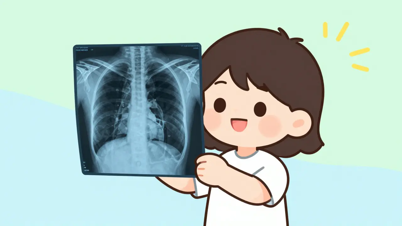
How doctors diagnose pneumothorax
Quick and accurate diagnosis saves lives. Here's what happens in the ER:
| Method | Sensitivity | Specificity | Time to Result |
|---|---|---|---|
| Chest X-ray | 85-94% | 90-95% | 15-30 minutes |
| E-FAST ultrasoundpoint-of-care ultrasound using the "lung point" sign | 94% | 100% | 5-10 minutes |
| CT scan | 100% | 100% | 30-60 minutes |
Chest X-rays are the first-line test but miss up to 30% of cases in supine trauma patients. That's why E-FAST ultrasound has become essential in emergency settings. A 2022 study in the Journal of Emergency Medicine showed experienced providers detect pneumothorax with 94.3% accuracy using this method. For stable patients with unclear X-rays, CT scans provide definitive answers but take longer to arrange.
Emergency treatment protocols
Doctors follow strict guidelines based on severity:
- Tension pneumothorax: Immediate needle decompression within 2 minutes. A 14-gauge needle is inserted above the third rib in the midclavicular line to release trapped air. This is a life-saving procedure that stabilizes patients before surgery.
- Large pneumothorax (over 30% collapse): Chest tube insertion within 30 minutes. A 28F tube is placed between ribs to drain air. Success rates are 92%, but 15-30% of cases develop complications like infection.
- Small primary pneumothorax (under 2 cm rim): Observation with supplemental oxygen. This allows air to be reabsorbed naturally. The Pneumostat trial showed 82% resolve within 14 days without intervention.
The American Heart Association's 2023 guidelines mandate that unstable patients get needle decompression before imaging. Waiting for X-rays in these cases can be fatal. For stable patients, oxygen therapy speeds recovery-10-15 L/min via non-rebreather mask increases air resorption from 1.25% to 4.2% per hour.
Recovery and prevention
After treatment, follow these evidence-based steps:
- Quit smoking: This is the single most effective prevention. The 2022 Tobacco and Respiratory Outcomes study found quitting reduces recurrence risk by 77% within a year.
- Avoid air travel: Wait 2-3 weeks after full resolution per FAA guidelines. Flying too soon can cause re-collapse due to cabin pressure changes.
- Never scuba dive: Without surgical intervention, diving carries a 12.3% recurrence rate. The Diving and Hyperbaric Medicine study confirms this risk is permanent for untreated pneumothorax.
- Follow-up X-ray: Get checked at 4-6 weeks. Studies show 8% of patients develop delayed complications without monitoring.
For recurrent cases, surgery is often needed. Video-assisted thoracoscopic surgery (VATS) has a 95% success rate at 1 year but costs about $18,500 in the U.S. according to 2023 Medicare data. The American Thoracic Society recommends surgical repair after a second ipsilateral episode due to the 62% recurrence rate after two episodes.
Frequently Asked Questions
Can a pneumothorax happen to someone with no lung problems?
Yes. Primary spontaneous pneumothorax occurs in healthy individuals without known lung disease. It's most common in tall, thin young men aged 20-40. About 7.4-18 per 100,000 people experience this annually, according to the American Thoracic Society's 2023 guidelines.
How long does it take for a collapsed lung to heal?
Small primary pneumothoraces often resolve in 7-10 days with oxygen therapy. Larger cases requiring chest tubes typically need 3-5 days of drainage. Full lung re-expansion usually takes 2-4 weeks. However, the British Thoracic Society warns that patients with secondary pneumothorax (due to underlying lung disease) may have longer recovery times and higher complication risks.
Is a collapsed lung dangerous?
Yes, especially if it progresses to tension pneumothorax. While primary cases have only 0.16% 30-day mortality, secondary cases in older patients with lung disease have a 16.2% mortality rate. The New England Journal of Medicine emphasizes that time to treatment is critical-each 30-minute delay increases complication risk by 7.2%.
Can I exercise after a pneumothorax?
Avoid strenuous activity for 2-3 weeks after full resolution. Light walking is usually fine once you're home from the hospital. The American Lung Association recommends waiting until your doctor confirms complete healing on X-ray before returning to sports or heavy lifting. Diving and high-altitude activities require surgical intervention to be safe.
What's the difference between primary and secondary pneumothorax?
Primary pneumothorax occurs in people without underlying lung disease-typically young, healthy individuals. Secondary pneumothorax happens when lung disease like COPD, cystic fibrosis, or pneumonia causes the collapse. Secondary cases are more severe, with higher recurrence rates (43-52% within a year) and significantly higher mortality compared to primary cases.
Savannah Edwards
February 7, 2026 AT 23:35I had a pneumothorax last year while hiking in Colorado. It was terrifying-sharp pain in my chest, couldn't catch my breath.
I remember thinking it was just a muscle cramp until the pain got worse. The ER docs explained it's when air leaks into the pleural space.
They did a chest X-ray and found a 20% collapse. They put in a chest tube, which was uncomfortable but saved my life.
What's scary is how quickly it can escalate to tension pneumothorax. I didn't know about the 'lung point' sign in ultrasound until later.
The American Thoracic Society stats are spot on-small collapses can sometimes be managed with oxygen, but if you're symptomatic, don't wait.
I quit smoking after this and have been fine for a year. It's a reminder to take chest pain seriously, no matter how 'healthy' you think you are.
For example, the study mentioned in the article about oxygen therapy speeding up resorption-10-15 L/min via non-rebreather mask increases it from 1.25% to 4.2% per hour. That's a huge difference.
Also, the FAA guidelines about waiting 2-3 weeks before flying-important to follow that.
I had a follow-up X-ray at 6 weeks and they confirmed full healing.
If you're young and tall like me, it's more common, but it can happen to anyone.
The key is recognizing the symptoms early. Sharp chest pain that worsens with breathing is a red flag.
Mayank Dobhal
February 9, 2026 AT 04:31Had a friend with pneumothorax. Was playing basketball, felt sharp pain, couldn't breathe. Called 911 right away. They did a chest tube and he's fine now. Don't ignore chest pain-it's life-threatening. Time is critical.
Marcus Jackson
February 9, 2026 AT 09:09The article says tension pneumothorax requires needle decompression in 2 minutes. But most people don't know how to do that. It's not like you can just stick a needle in yourself. ERs need to be ready.
Also, the E-FAST ultrasound is way better than X-rays for quick diagnosis. Should be standard in ambulances. Maybe the article should mention that.
Natasha Bhala
February 11, 2026 AT 05:10Call 911 for chest pain and shortness of breath.
Jesse Lord
February 11, 2026 AT 22:38I really appreciate you sharing this its important to know the symptoms. I work in healthcare and see how fast things can go wrong. Always better to act quickly. Thanks for the info
Gouris Patnaik
February 13, 2026 AT 07:52In India healthcare is far superior. We handle pneumothorax cases with precision. US system is broken. People there ignore symptoms until it's too late. Shameful.
Ashley Hutchins
February 14, 2026 AT 20:29I think people who ignore chest pain deserve what they get. Smoking is the main cause. If you dont quit youre asking for trouble. No sympathy for those who dont take responsibility
Lakisha Sarbah
February 15, 2026 AT 14:38good point about E-FAST. ive seen it used in my hospital. much faster than X-rays. but it takes training. maybe more paramedics should learn it
AMIT JINDAL
February 16, 2026 AT 12:57Hey there! So glad you shared your experience! 😊 As someone who's been through multiple medical procedures, I can say that the 'lung point' sign in ultrasound is game-changing. It's like the medical community has finally caught up with modern tech. I mean, X-rays are outdated for this! 😂 Also, the oxygen therapy stats are spot on. But honestly, the real issue is smoking-people just don't quit. 🤦♂️ If you're from a 'healthy' background, you should know better. Anyway, stay safe and keep that lung healthy! 💪
Ariel Edmisten
February 17, 2026 AT 10:22you're right. acting fast saves lives. im glad er's are getting better at this. keep spreading awareness
Niel Amstrong Stein
February 19, 2026 AT 03:51Interesting perspective from India. Healthcare systems vary globally, but the core issue is timely intervention. 🌍 Whether in US or India, recognizing symptoms early is key. Maybe we should focus on education rather than blaming systems. Peace out! ✌️
Paula Sa
February 20, 2026 AT 16:30i see where you're coming from about smoking but it's not that simple. addiction is a disease. maybe instead of judgment we should support people in quitting. that's more effective. just saying